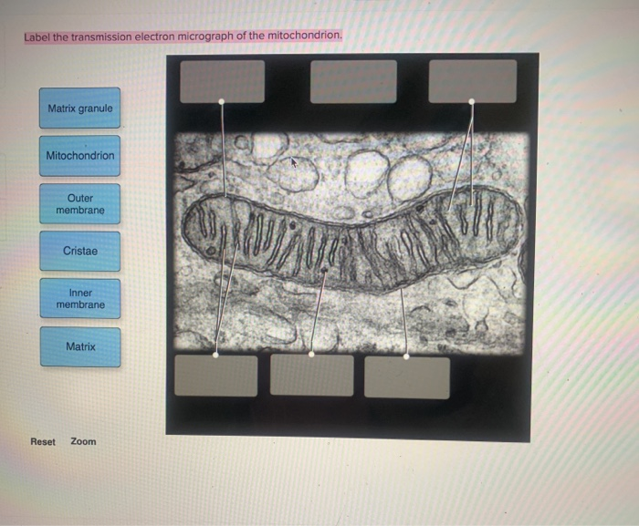
42 transmission electron micrograph labeled
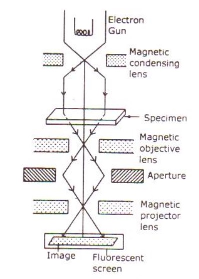
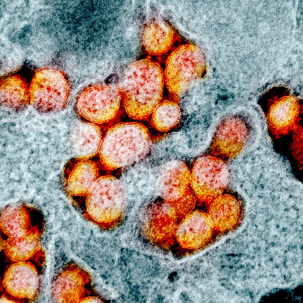
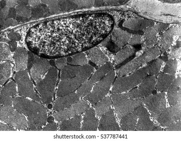
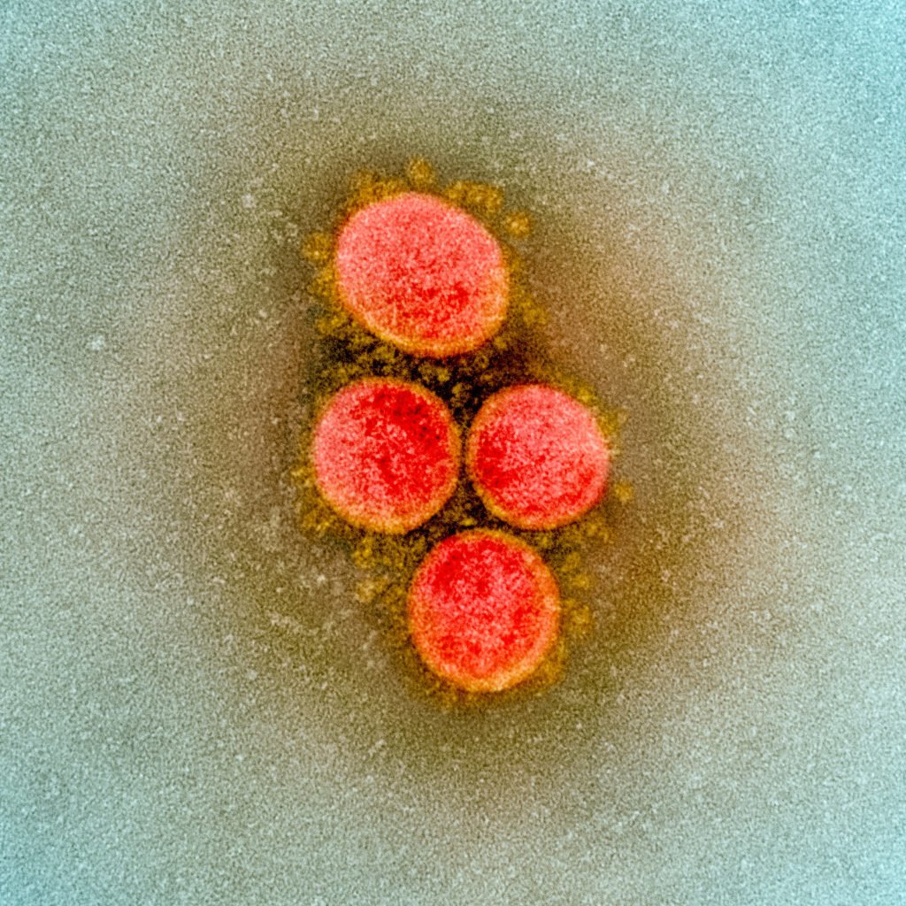
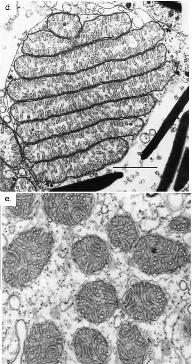
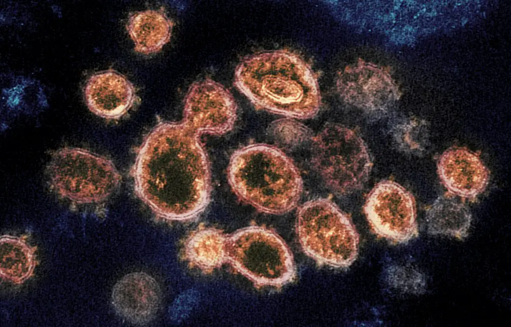
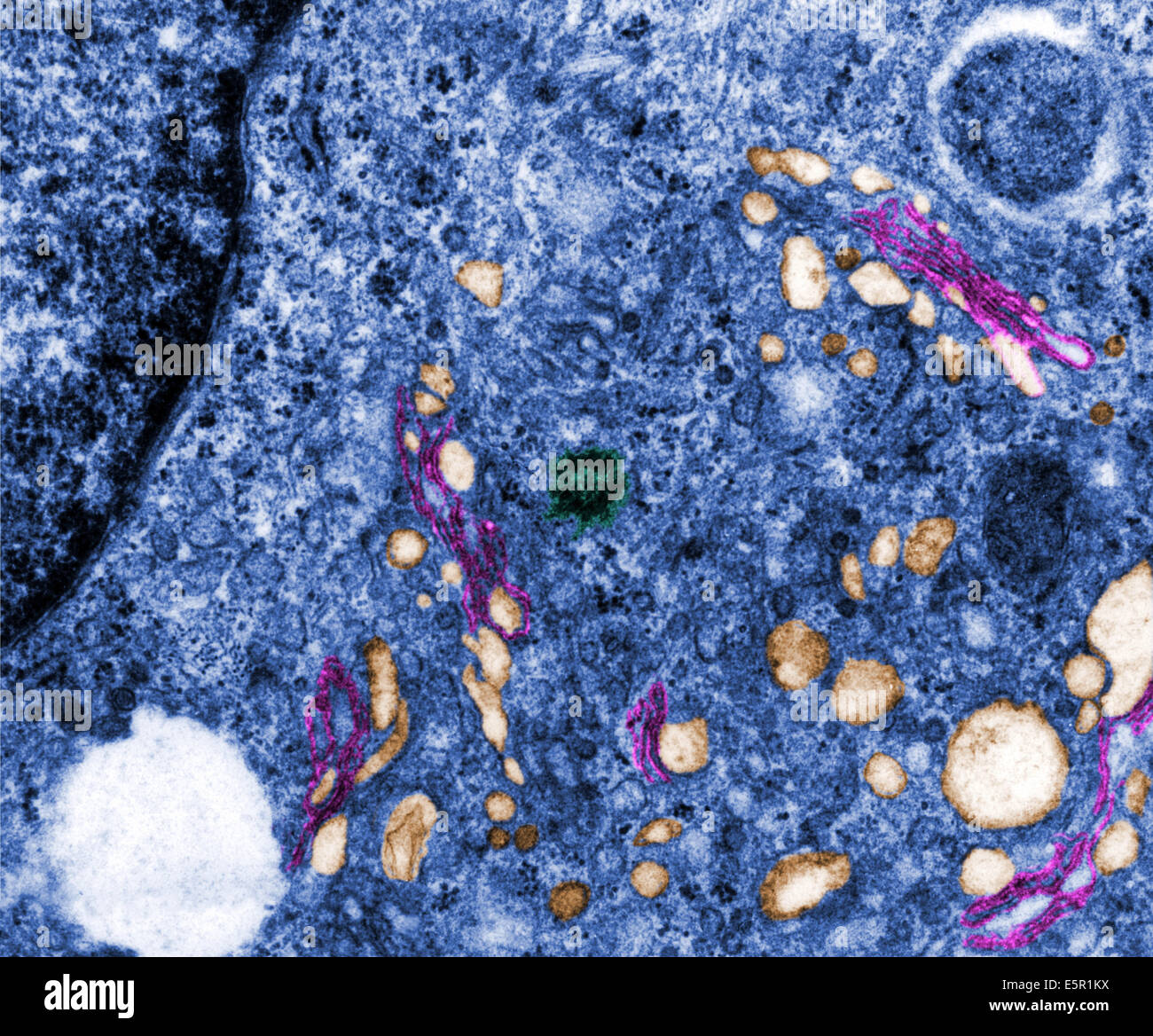
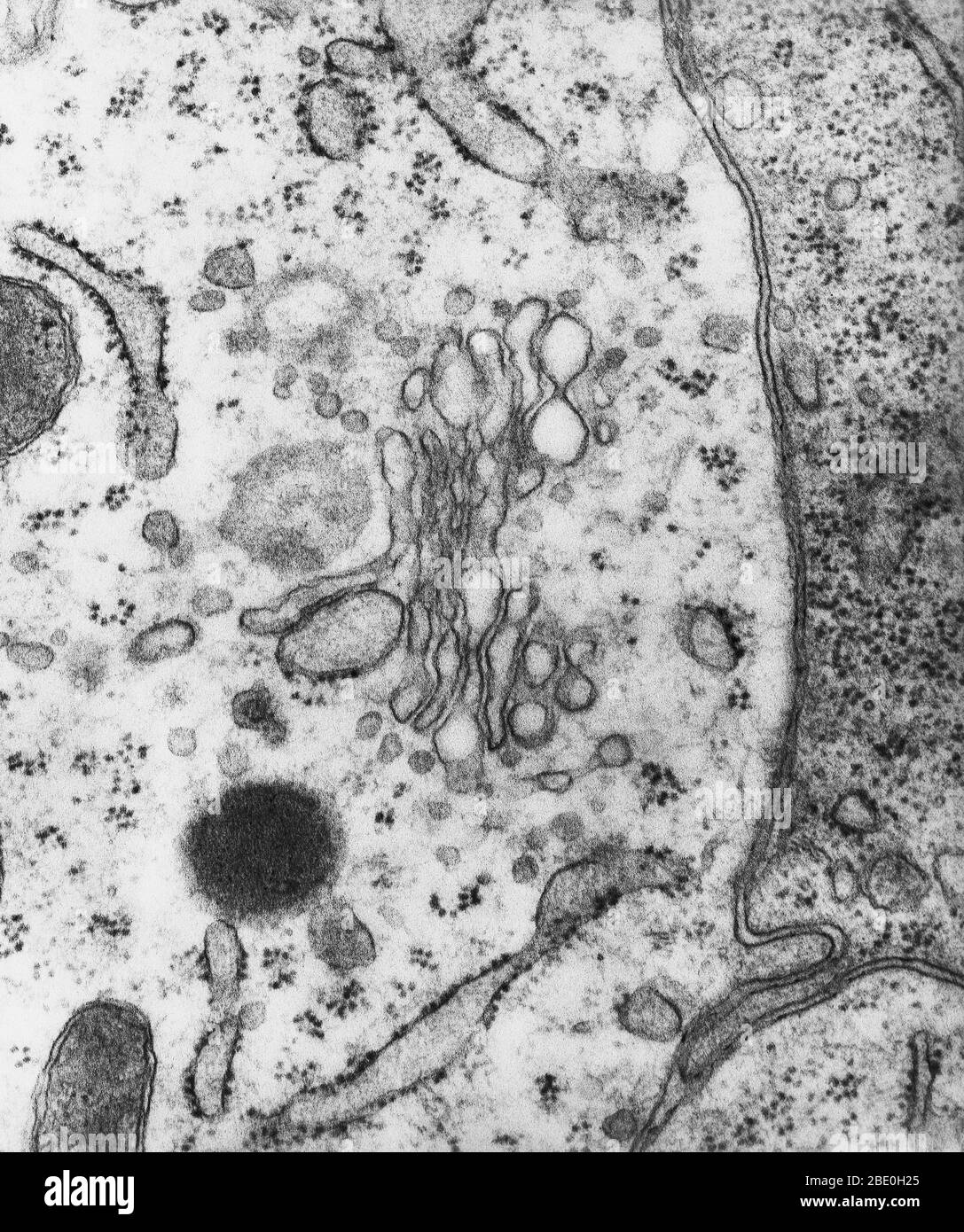
Transmission Electron Microscope (With Diagram) - Biology Discussion The final image in a TEM is known as transmission electron micrograph. The salts of some heavy metals, e.g., lead; osmium, tungsten and uranium are often used for staining. These heavy metal stains are used to increase the contrast between ultra structures and the background. › media › subtopicImage Library | CDC Online Newsroom | CDC Note the spikes that adorn the outer surface of the virus, which impart the look of a corona surrounding the virion, when viewed electron microscopically. In this view, the protein particles E, S, and M, also located on the outer surface of the particle, have all been labeled as well.
› cryptosporidiumCryptosporidium - Morphology, Microscopy, Tests, Infection ... · Antibodies labeled with enzyme reporters - Currently, some of the kits available for antigen detection using antibodies labeled with enzyme reporters include Enzyme Immunoassay (EIA) and Immunochromatographic (IC). Using EIA kits (in microplate format), which has 93 to 100 percent sensitivity and specificity; it's possible to detect ...
Transmission electron micrograph labeled
rsscience.com › cell-membraneCell membrane - definition, structure, function, and biology T-tubules permit the rapid transmission of the action potential into the cell, allowing heart muscle cells to contract more forcefully. Photo source: wiki [In this figure] Electron micrograph showing the surface of endothelial cells’ membrane coated with a thick layer of carbohydrate components, called glycocalyx . Microscope Types (with labeled diagrams) and Functions The shorter wavelength of electrons compared to visible light photons helps the observer achieve a very high resolving power compared to normal microscopes thereby aiding observers to see very tiny objects clearly. Electron microscope labeled diagram The different types of electron microscopes are: Transmission Electron Microscope Solved Label the transmission electron micrograph of the - Chegg Transcribed image text: Label the transmission electron micrograph of the cell. 0 Nucleus rences Mitochondrion Heterochromatin Peroxisome Vesicle ULAR bumit Click and drag each label into the correct category to indicate whether it pertains to the cytoplasm or the plasma membrane.
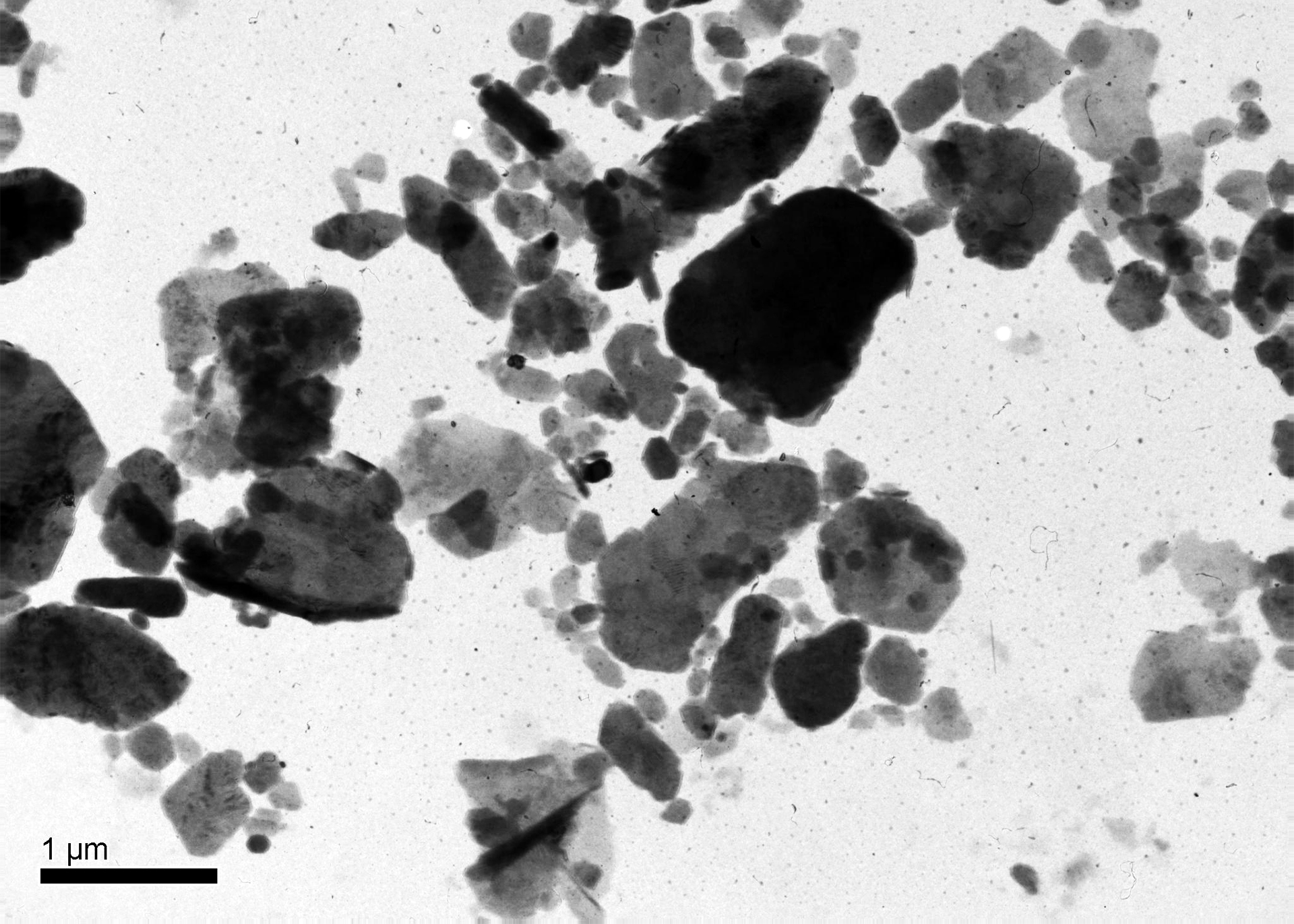
Transmission electron micrograph labeled. Label This Transmission Electron Micrograph - Kaiden Brown Label this transmission electron micrograph of relaxed sarcomeres by clicking and dragging the labels to the correct location . Transmission electron microscopy (tem) is one of the oldest technologies and still. Molecular labeling for correlative microscopy: Fluorescence microscopy in combination with tem and an ion beam analysis (iba, which ... Label This Transmission Electron Micrograph Of A Relaxed ... - Blogger Label this transmission electron micrograph of relaxed sarcomeres by clicking and dragging the labels to the correct location . Label the following image using the terms provided. Note how the sarcomeres are extended to only approximately 120 % . IMG_2132 - FIGURES Label this transmission electron from Transmission Electron Micrographs of Negatively Stained Salmonella ... This low magnification micrograph depicts a single bacterium possessing multiple flagella; the arrow indicates one of these flagella. Protein aggregates from the media are visible as light-colored circles. Bar = 1 µm. Figure 2: Negatively Stained Transmission Electron Micrograph of Salmonella typhimurium (Labeled view). Transmission Electron Microscope (TEM) - Uses, Advantages and Disadvantages A Transmission Electron Microscope is an impressive instrument with a number of advantages such as: TEMs offer the most powerful magnification, potentially over one million times or more. TEMs have a wide-range of applications and can be utilized in a variety of different scientific, educational and industrial fields.
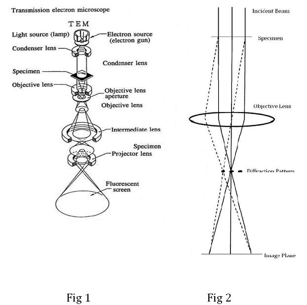
Transmission Electron Microscope (TEM)- Definition, Principle, Images Parts of Transmission Electron Microscope (TEM) Their working mechanism is enabled by the high-resolution power they produce which allows it to be used in a wide variety of fields. It has three working parts which include: Electron gun Image producing system Image recording system Electron gun Electron Microscope-Definition, Principle, Types, Uses, Labeled Diagram There are two varieties of electron microscope, each having a unique way of working: 1. Transmission Electron Microscope (TEM) Thin specimens that allow electrons to flow through and produce a picture are seen using a transmission electron microscope. The standard (compound) light microscope and the TEM are similar in many aspects. Assignment 6, page 2 - North Carolina State University The micrograph is displayed as if using a "virtual electron microscope", so you will need to magnify the image and move to a region that contains the clearest view of chloroplast internal structures. Perform a screen capture of the chloroplast, then label: 1) a thylakoid, 2) a granum, 3) the stroma, and 4) the outer chloroplast membrane. Submit ... Label This Transmission Electron Micrograph / Microscopy Innovations ... Label the transmission electron micrograph of the nucleus. Transmission electron micrographs of hela cell sections labeled in . Label the transmission electron micrograph of the nucleus. Fluorescence microscopy in combination with tem and an ion beam analysis (iba, which allows the evaluation of the chemical elemental distribution) has allowed .
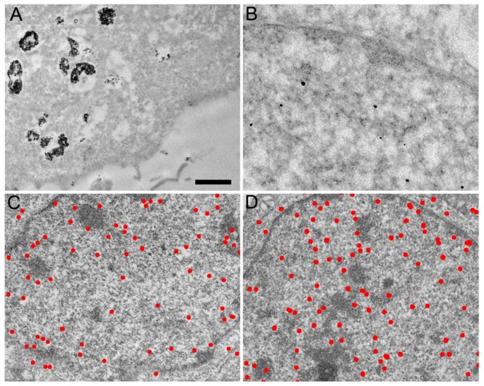
Transmission electron micrograph (TEM) identifying immunogold labeled ... Transmission electron micrograph (TEM) identifying immunogold labeled ESR1 and caveolin-1 proteins in the uterine artery endothelial cells derived from the pregnant state (P-UAEC). (A) IgG control... Concatenated metallothionein as a clonable gold label for electron ... Visualization of gold-labeled fusion proteins by scanning electron microscopy required one copy of metallothionein while transmission electron microscopy required two copies. Images of frozen-hydrated samples of simple complexes made with anti-MBP antibodies hint at the usefulness of this method. Publication types Transmission Electron Microscopy (TEM) - Warwick TEM. The transmission electron microscope is a very powerful tool for material science. A high energy beam of electrons is shone through a very thin sample, and the interactions between the electrons and the atoms can be used to observe features such as the crystal structure and features in the structure like dislocations and grain boundaries. Electron Micrographs - University of Oklahoma Health Sciences Center Electron Micrographs. Below is a collection of electron micrographs with labelled subcellular structures that you should be able to identify. Also, be sure to observe any electron micrographs which are made available in the laboratory by the instructor. You should concentrate on the similarities in form that permit identification of the ...
Transmission Electron microscope - Principle, Construction ...
elifesciences.org › articles › 80047Automated systematic evaluation of cryo-EM specimens with ... Aug 23, 2022 · The complexity of a screening workflow depends on several factors including the instrument used and the type of specimen. Here, we describe the extended operation of a microscope furnished with an autoloader device and loaded with frozen hydrated targets for SPA, which are prepared on a micropatterned holey substrate or continuous carbon (Figure 1 and Figure 1—figure supplement 1).
Live cell immunogold labelling of RNA polymerase II ...
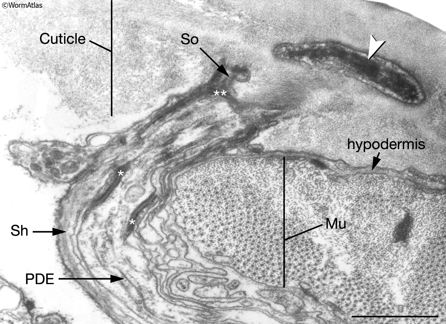
Transmission Electron Microscopy - an overview | ScienceDirect Topics Transmission Electron Microscopy. Transmission electron microscopy (TEM) is a powerful tool for examination of the microanatomy of biological tissues, cells and organisms including nematodes. ... Clusters of wheat germ agglutinin-labeled gold granules were located all over the surface of milk fat globules using SEM, suggesting the presence of N ...

4.4 The Endomembrane System and Proteins | Texas Gateway
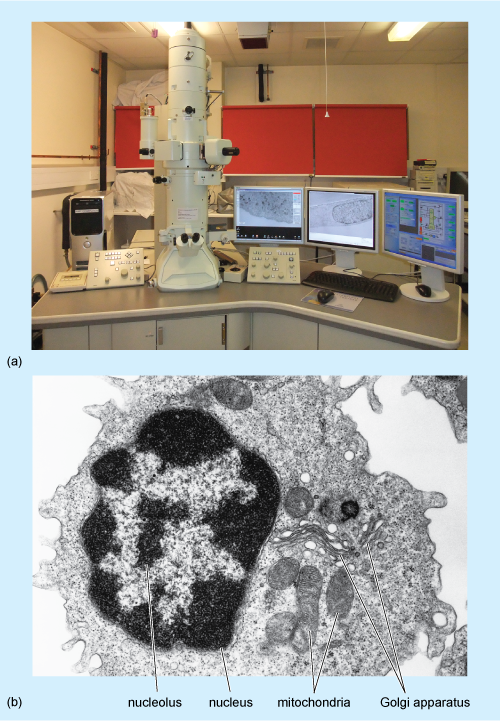
Electron Microscope- Definition, Principle, Types, Uses, Labeled Diagram There are two types of electron microscopes, with different operating styles: 1. Transmission Electron Microscope (TEM) The transmission electron microscope is used to view thin specimens through which electrons can pass generating a projection image. The TEM is analogous in many ways to the conventional (compound) light microscope.
Transmission electron microscopy cells hi-res stock ...
Transmission electron microscopy of macrophages and mic | Open-i Transmission electron microscopy of macrophages and microglial cells after 4 day feeding with HNE-modified ROSs. A: Electron micrograph of microglial cells afte
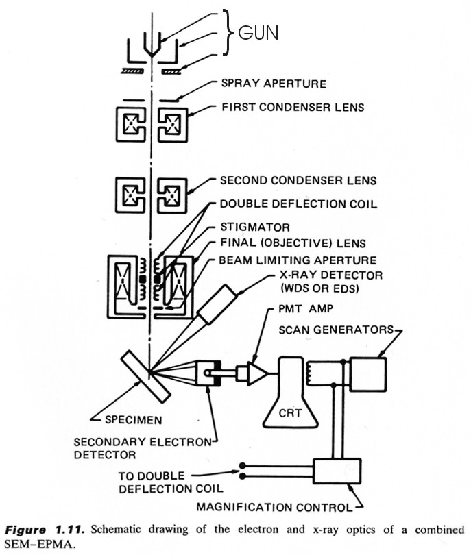
Scanning Electron Microscopy (SEM)
link.aps.org › doi › 10Rev. Mod. Phys. 81, 109 (2009) - The electronic properties of ... Jan 14, 2009 · (a) Bright-field transmission-electron-microscope image of a graphene membrane. Its central part (homogeneous and featureless region) is monolayer graphene. Adapted from 263. (b) Despite only one atom thick, graphene remains a perfect crystal at this atomic resolution. The image is obtained in a scanning transmission electron microscope.
Transmission electron microscopy (TEM) micrographs of E. coli ...
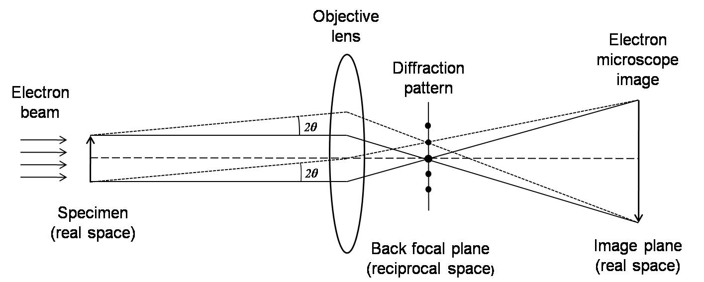
Transmission electron microscopy - Wikipedia Transmission electron microscopy (TEM) is a microscopy technique in which a beam of electrons is transmitted through a specimen to form an image. The specimen is most often an ultrathin section less than 100 nm thick or a suspension on a grid. An image is formed from the interaction of the electrons with the sample as the beam is transmitted through the specimen.

Solved Label the transmission electron micrograph based on ...
Transmission Electron Micrograph of transfected HL-1 cells labeled for ... Transmission Electron Micrograph of transfected HL-1 cells labeled for TMEM43 with immunogold. A and B. Single immunogold labeling experiments used 15 nm gold particles to label GFP. A....

Chapter 14 & 15 Flashcards Flashcards | Quizlet
Transmission Electron Microscopy - Penn State College of Medicine Research The JEOL 1400 TEM (Room C1727) is capable of generating ultra-structural nanoscale images from fixed cell/tissue samples or multiplexed immune-labeled samples. Computer-controlled operations Resolution up to 3 Angstroms Magnification up to 370,000X Capable of collecting data suitable for 3D reconstructions of negative-stained samples
Transmission Electron Microscope (TEM)- Definition, Principle ...
PDF Identifying Organelles from an Electron Micrograph The photograph shown below details chloroplast structure as viewed with a transmission electron microscope Courtesy of Dr. Julian Thorpe - EM & FACS Lab, Biological Sciences University Of Sussex A single Granum Chloroplast envelope visible as two membranes Stroma containing numerous small ribosomes Lamellae connecting different grana

Scanning Transmission Electron Microscopy - an overview ...
The Transmission Electron Microscope | CCBER - UC Santa Barbara Transmission electron microscopes (TEM) are microscopes that use a particle beam of electrons to visualize specimens and generate a highly-magnified image. TEMs can magnify objects up to 2 million times. In order to get a better idea of just how small that is, think of how small a cell is. It is no wonder TEMs have become so valuable within the ...
CcFIG 5 Legend
Scanning transmission electron microscopy/energy-dispersive ... in an attempt to compare the morphology of the dentin adhesive interface and the wetting and penetration of the adhesive in relation to the dentin surface, we studied four dentin adhesive systems using scanning transmission electron microscopy (stem) and energy-dispersive spectroscopy (eds). 2-hydroxyethylmethacrylate (hema), a monomer common to …
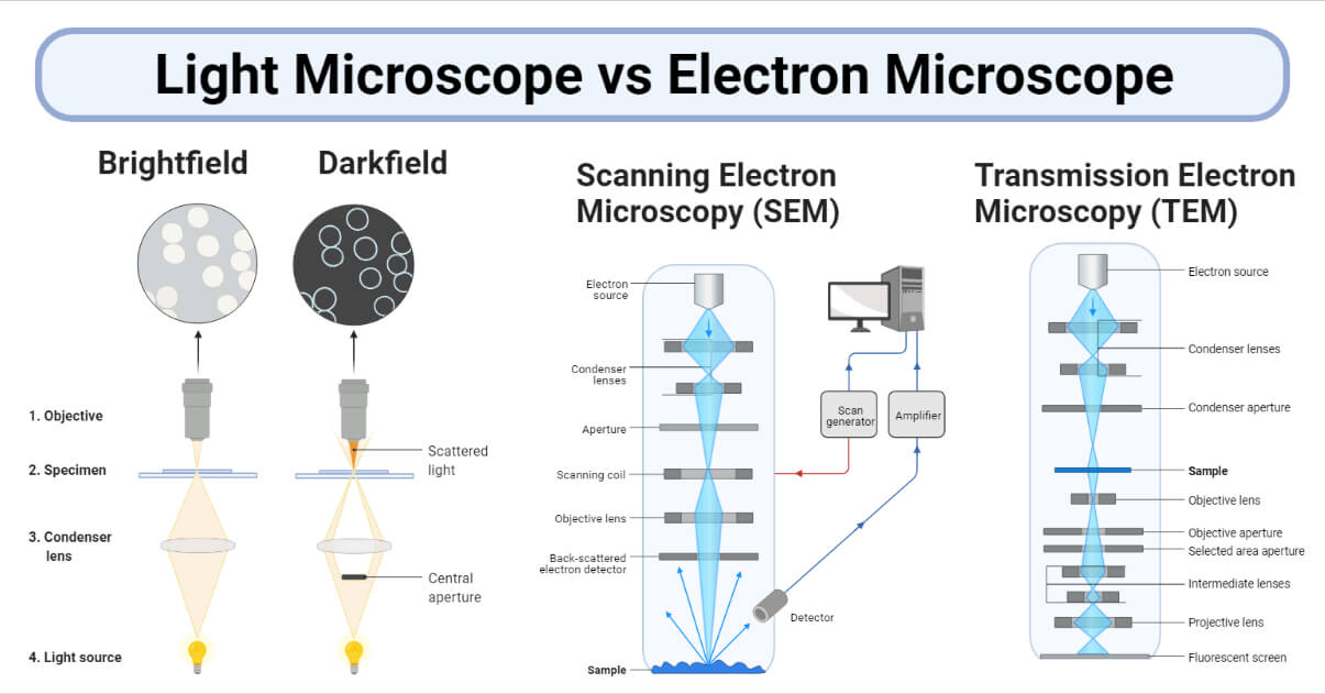
Light Microscope vs Electron Microscope- 36 Major Differences
en.wikipedia.org › wiki › SmallpoxSmallpox - Wikipedia The envelope was labeled as containing scabs from a vaccination and gave scientists at the CDC an opportunity to study the history of smallpox vaccination in the United States. On July 1, 2014, six sealed glass vials of smallpox dated 1954, along with sample vials of other pathogens, were discovered in a cold storage room in an FDA laboratory ...

Cell Micrographs | BioNinja
Transmission electron microscopy DNA sequencing - Google Transmission electron microscopy DNA sequencing is a single-molecule sequencing technology that uses transmission electron microscopy techniques.The method was conceived and developed in the 1960s and 70s, but lost favor when the extent of damage to the sample was recognized. DNA is visible under the electron microscope; however, it must be labeled with heavy atoms so that the DNA bases can be ...

Transmission electron microscopy (TEM). Ten-nanometre gold ...
en.wikipedia.org › wiki › History_of_cell_membraneHistory of cell membrane theory - Wikipedia A more direct investigation of the membrane was made possible through the use of electron microscopy in the late 1950s. After staining with heavy metal labels, Sjöstrand et al. noted two thin dark bands separated by a light region, which they incorrectly interpreted as a single molecular layer of protein. A more accurate interpretation was ...
Lecture 31 Light Microscopy
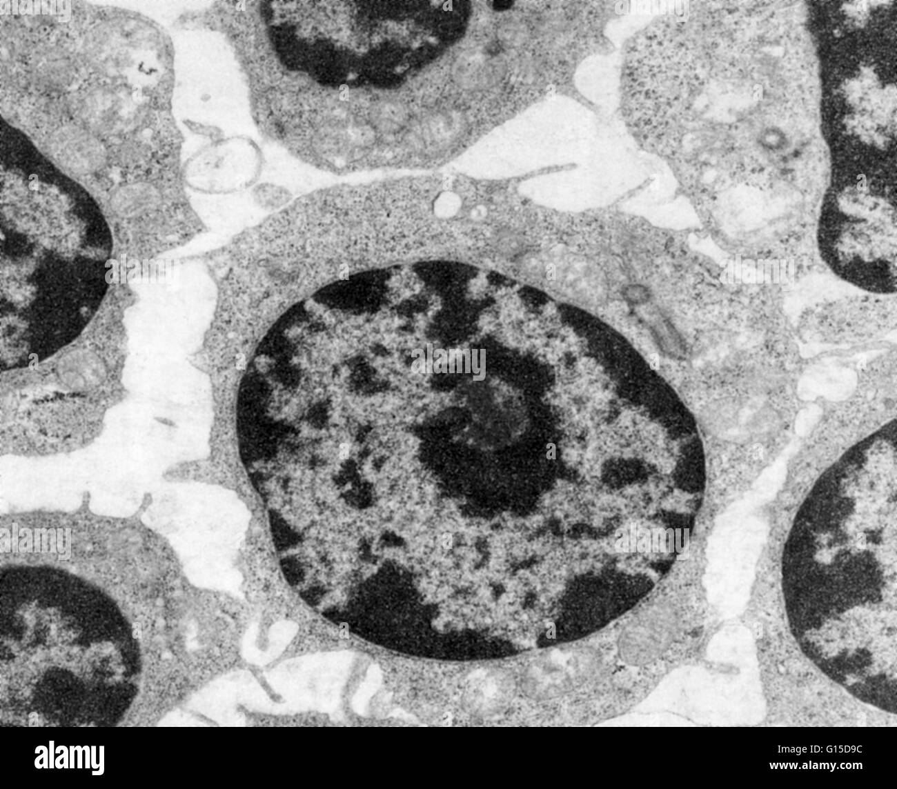
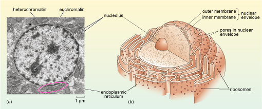
Labeling the Cell Flashcards | Quizlet Label the transmission electron micrograph of the nucleus. membrane bound organelles golgi apparatus, mitochondrion, lysosome, peroxisome, rough endoplasmic reticulum nonmembrane bound organelles ribosomes, centrosome, proteasomes cytoskeleton includes microfilaments, intermediate filaments, microtubules Identify the highlighted structures
8.2: Transmission Electron Microscopy - Chemistry LibreTexts
Solved Label this transmission electron micrograph of - Chegg Anatomy and Physiology questions and answers Label this transmission electron micrograph of relaxed sarcomeres by clicking and dragging the labels to the correct location Sarcamere 1 band (light) Z disc Mline Aband (dark) H zone
586 Transmission electron micrograph Images, Stock Photos ...
Transmission electron microscopy DNA sequencing - Wikipedia Transmission electron microscopy (TEM) produces high magnification, high resolution images by passing a beam of electrons through a very thin sample. Whereas atomic resolution has been demonstrated with conventional TEM, further improvement in spatial resolution requires correcting the spherical and chromatic aberrations of the microscope lenses.
IntroFIG 4B Legend
Solved Label the transmission electron micrograph of the - Chegg Transcribed image text: Label the transmission electron micrograph of the cell. 0 Nucleus rences Mitochondrion Heterochromatin Peroxisome Vesicle ULAR bumit Click and drag each label into the correct category to indicate whether it pertains to the cytoplasm or the plasma membrane.
Transmission Electron Microscope (TEM)- Definition, Principle ...
Microscope Types (with labeled diagrams) and Functions The shorter wavelength of electrons compared to visible light photons helps the observer achieve a very high resolving power compared to normal microscopes thereby aiding observers to see very tiny objects clearly. Electron microscope labeled diagram The different types of electron microscopes are: Transmission Electron Microscope
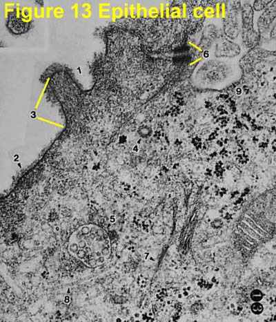
Electron Micrographs
rsscience.com › cell-membraneCell membrane - definition, structure, function, and biology T-tubules permit the rapid transmission of the action potential into the cell, allowing heart muscle cells to contract more forcefully. Photo source: wiki [In this figure] Electron micrograph showing the surface of endothelial cells’ membrane coated with a thick layer of carbohydrate components, called glycocalyx .

Transmission electron micrograph of turkey spermatozoa ...

Solved Mitochondrion Nucleus Vesicle Peroxisome | Chegg.com
Transmission electron micrographs of mitochondria, site of ...
Transmission Electron Microscopy (TEM)
What is a diagram of a plant and animal cell under an ...

Transmission electron microscope for USPIO-labeled cells ...
Transmission Electron Microscope: Definition, Parts, Working ...
A tour of the cell: View as single page
Transmission electron micrograph (TEM) of the Golgi apparatus ...
What is Transmission Electron Microscopy?
Solved Label the transmission electron micrograph of the ...
A tour of the cell: View as single page

Transmission electron microscopy (TEM) of graphene ...
TEM monsters
8.2: Transmission Electron Microscopy - Chemistry LibreTexts
Immunogold labelling - Wikipedia
Transmission electron microscopy cells hi-res stock ...
ExcFIG 4 Legend
Transmission electron microscopy cells hi-res stock ...

Transmission electron micrograph of a doublelabeled section ...
Microscopy Innovations | Transmission electron microscopy (TEM)

The Transmission Electron Microscope | CCBER
Post a Comment for "42 transmission electron micrograph labeled"