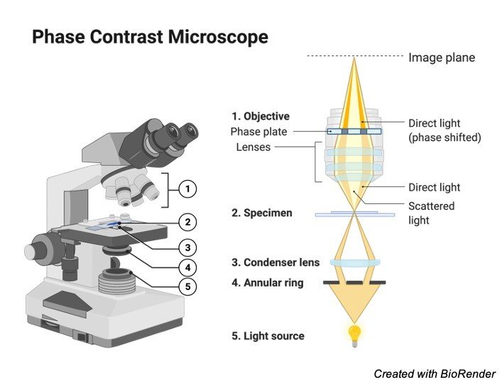
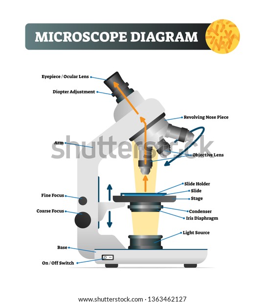
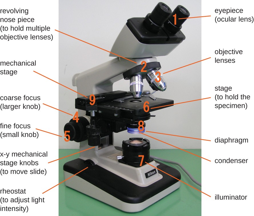
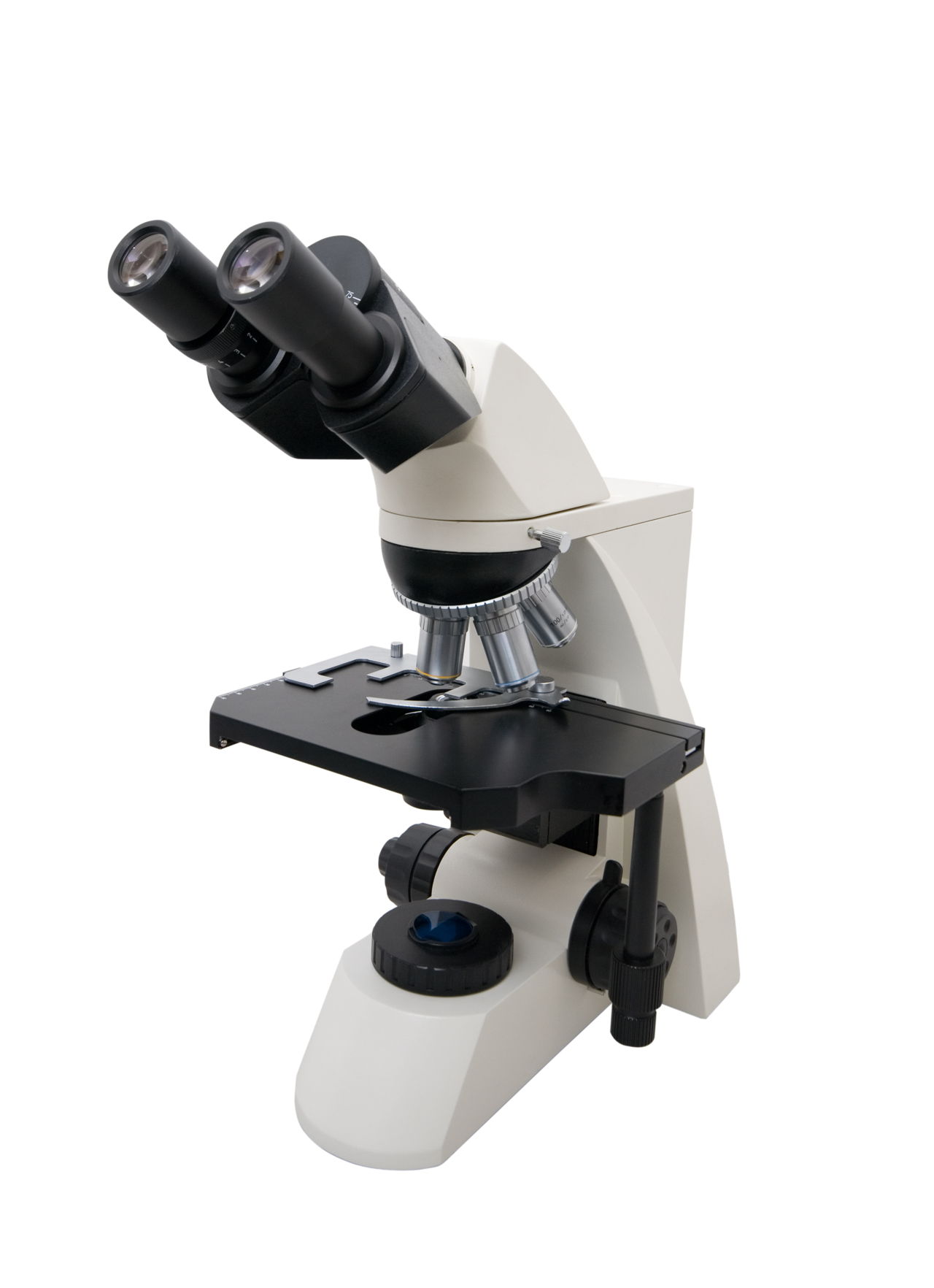
40 labeled diagram of microscope
Looking at the Structure of Cells in the Microscope A typical animal cell is 10–20 μm in diameter, which is about one-fifth the size of the smallest particle visible to the naked eye. It was not until good light microscopes became available in the early part of the nineteenth century that all plant and animal tissues were discovered to be aggregates of individual cells. This discovery, proposed as the cell doctrine by Schleiden and … Cat Skeleton Anatomy with Labeled Diagram - AnatomyLearner May 29, 2021 · Cat skeleton anatomy labeled diagram. Now, I will show you all the bones from the cat skeleton with a diagram. If you find any mistakes in this cat anatomy labeled diagram, please let me know. I hope this cat skeletal system anatomy labeled diagram might help you understand and identify all the cat’s bones.
Dog Skeleton Anatomy with Labeled Diagram - The Place to ... Dec 31, 2021 · Here, in the dog skeleton labeled diagram, I tried to show you the different segments of the forelimb, hindlimb with their bones. Again, I tried to show you all the bones from the vertebrae column of a dog skeleton. In addition, in the diagram, you will find a few identified skull bones. The sternum and the ribs are also identified in the dog ...
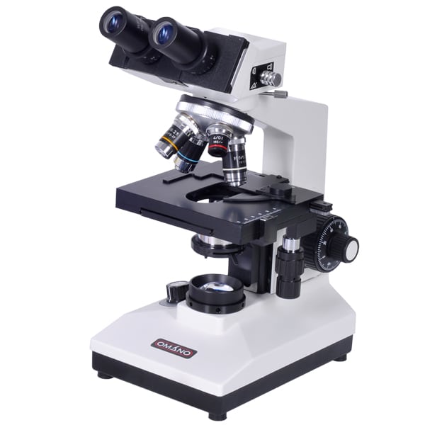
Labeled diagram of microscope
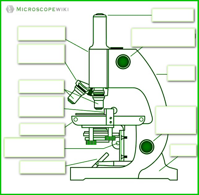
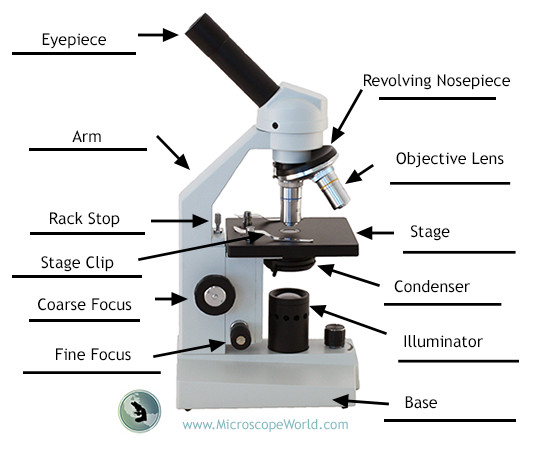
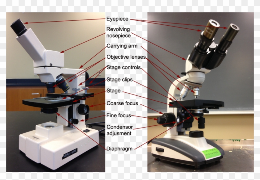
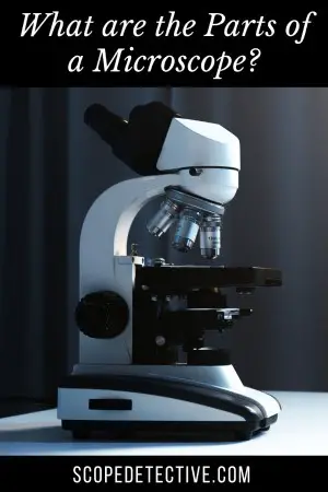
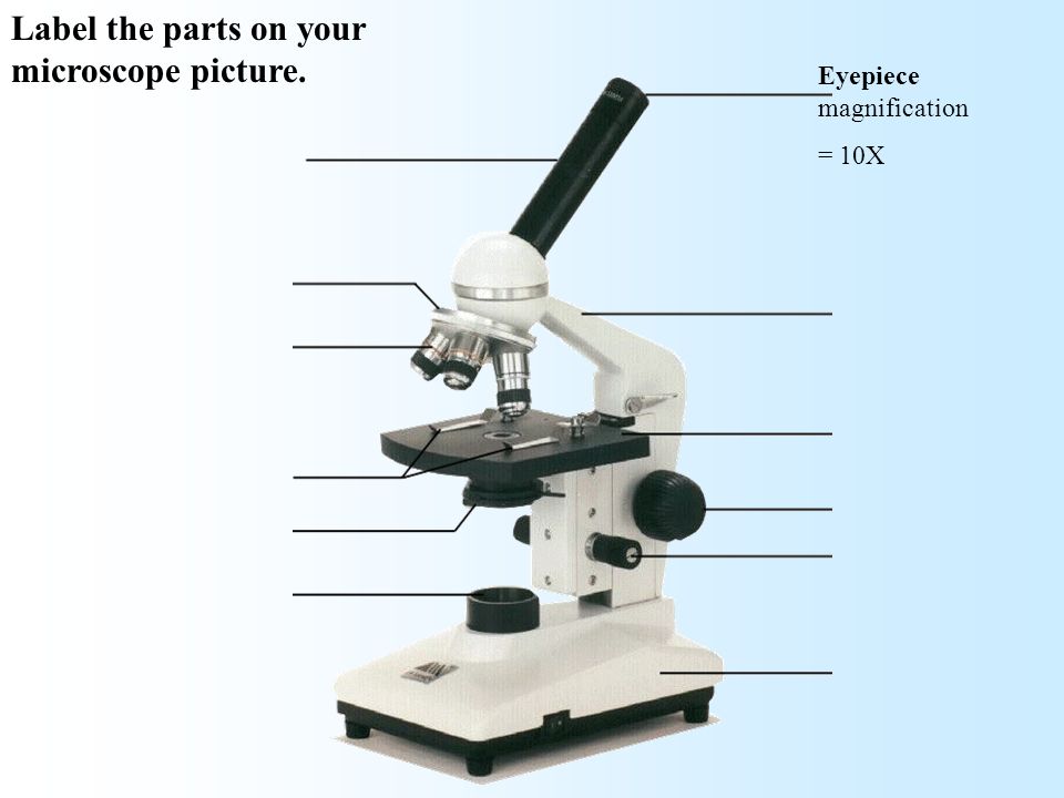
Microscope, Microscope Parts, Labeled Diagram, and Functions 19/01/2022 · Revolving Nosepiece or Turret: Turret is the part of the microscope that holds two or multiple objective lenses and helps to rotate objective lenses and also helps to easily change power. Objective Lenses: Three are 3 or 4 objective lenses on a microscope. The objective lenses almost always consist of 4x, 10x, 40x and 100x powers. The most common eyepiece … Compound Microscope- Definition, Labeled Diagram, Principle ... Apr 03, 2022 · Therefore, a microscope can be understood as an instrument to observe tiny elements. The optical microscope often referred to as the light microscope, is a type of microscope that uses visible light and a system of lenses to magnify images of small subjects. There are two basic types of optical microscopes: Simple microscopes; Compound microscopes Parts of a microscope with functions and labeled diagram Apr 19, 2022 · Figure: Diagram of parts of a microscope. There are three structural parts of the microscope i.e. head, base, and arm. Head – This is also known as the body. It carries the optical parts in the upper part of the microscope. Base – It acts as microscopes support. It also carries microscopic illuminators.
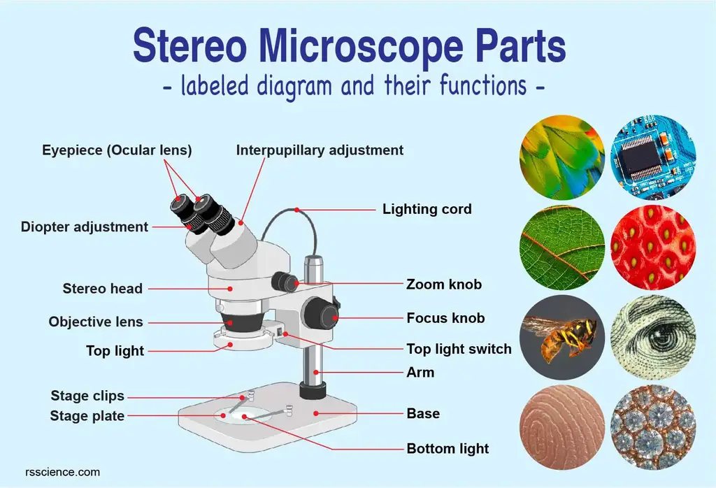
Labeled diagram of microscope. Microscope Parts and Functions Microscope Parts and Functions With Labeled Diagram and Functions How does a Compound Microscope Work?. Before exploring microscope parts and functions, you should probably understand that the compound light microscope is more complicated than just a microscope with more than one lens.. First, the purpose of a microscope is to magnify a small object or to … How does a Microscope work A simple microscope has one lens and is essentially a loupe or magnifying glass with a relatively high magnification. The basic modern microscope found in schools, hospitals, and research centers is a compound microscope which has a series of lenses to collect and focus the light transmitted through the specimen. What is a Stereo Microscope? - New York Microscope Company 11/05/2018 · Each part of a stereo microscope is labeled in the diagram below. This example is a typical classroom type stereo microscope with track stand and built-in illumination. Stereo Head: This is the moveable top portion of the microscope and the stereo head holds the two adjustable eyepieces. Ocular Lens ... Fluorescence microscope - Wikipedia A fluorescence microscope is an optical microscope that uses fluorescence instead of, or in addition to, scattering, reflection, and attenuation or absorption, to study the properties of organic or inorganic substances. "Fluorescence microscope" refers to any microscope that uses fluorescence to generate an image, whether it is a simple set up like an epifluorescence …
Parts of a microscope with functions and labeled diagram Apr 19, 2022 · Figure: Diagram of parts of a microscope. There are three structural parts of the microscope i.e. head, base, and arm. Head – This is also known as the body. It carries the optical parts in the upper part of the microscope. Base – It acts as microscopes support. It also carries microscopic illuminators. Compound Microscope- Definition, Labeled Diagram, Principle ... Apr 03, 2022 · Therefore, a microscope can be understood as an instrument to observe tiny elements. The optical microscope often referred to as the light microscope, is a type of microscope that uses visible light and a system of lenses to magnify images of small subjects. There are two basic types of optical microscopes: Simple microscopes; Compound microscopes Microscope, Microscope Parts, Labeled Diagram, and Functions 19/01/2022 · Revolving Nosepiece or Turret: Turret is the part of the microscope that holds two or multiple objective lenses and helps to rotate objective lenses and also helps to easily change power. Objective Lenses: Three are 3 or 4 objective lenses on a microscope. The objective lenses almost always consist of 4x, 10x, 40x and 100x powers. The most common eyepiece …















Post a Comment for "40 labeled diagram of microscope"