42 photomicrograph of thick skin
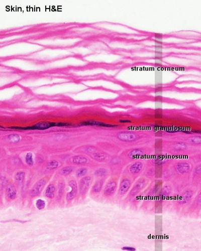
SKIN | The Big Picture: Histology | AccessBiomedical Science | McGraw ... A subcutaneous layer of loose connective tissue below the dermis that attaches the skin to underlying tissues. Skin contains various appendages derived from epidermis, including sweat glands, hair follicles and sebaceous glands. Skin is classified as either thick or thin. Thick skin is found on the palms and the soles. Low Magnification Micrograph Human Thin Skin Stock Photo ... - Shutterstock Download for free. Royalty-free stock photo ID: 1136062721. Low magnification micrograph of a human thin skin showing the epidermis and a very thick dermis, characteristic of this type of skin. Hematoxylin-eosin.
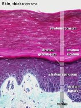
Skin: The Histology Guide Dermis: Thick skin has a thinner dermis than thin skin, and does not contain hairs, sebaceous glands, or apocrine sweat glands. Thick skin is only found in areas where there is a lot of abrasion - fingertips, palms and the soles of your feet. show labels This is a picture of an H&E stained section of the epidermis of thin skin.

Photomicrograph of thick skin
quizlet.com › 511891167 › bio-232-lab-midterm-flashBio 232 ~ Lab Midterm Flashcards - Quizlet Label the photomicrograph of thin skin. Place the following layers in order from superficial to deep. Fill-in the blanks with the appropriate tissue type being described. pubs.rsna.org › doi › fullPulmonary Tuberculosis: Role of Radiology in Diagnosis and ... Jan 11, 2017 · Tuberculosis is a public health problem worldwide, including in the United States—particularly among immunocompromised patients and other high-risk groups. Tuberculosis manifests in active and latent forms. Active disease can occur as primary tuberculosis, developing shortly after infection, or postprimary tuberculosis, developing after a long period of latent infection. Primary tuberculosis ... In the photomicrograph of a portion of thick skin - Course Hero Section Reference 1: Sec 5.1 Structure of the Skin. 33) In the photomicrograph of a portion of thick skin shown below, which layer is the stratum basale? a) Ab) B c) D d) Ee) F Answer: d. Difficulty: Medium Study Objective 1: SO 5.1 Describe the general structure of the skin.
Photomicrograph of thick skin. Skin overview 4 - Digital Histology Skin can be classified as either thick or thin, depending on the thickness of the epidermal layer. A diagrammatic representation of thin skin and a photomicrograph of a H&E stained section illustrate the reduced thickness of the strata in thin skin and the absence of stratum lucidum as a distinct layer. 400x. - Papillary layer of dermis. Solved Label the photomicrograph of thick skin | Chegg.com Label the photomicrograph of thick skin ; Question: Label the photomicrograph of thick skin . This problem has been solved! See the answer See the answer See the answer done loading. Show transcribed image text Expert Answer. Who are the experts? Experts are tested by Chegg as specialists in their subject area. We review their content and use ... photomicrograph of thick skin Diagram | Quizlet photomicrograph of thick skin Diagram | Quizlet photomicrograph of thick skin STUDY Learn Write Test PLAY Match Created by mckennawebber Terms in this set (7) epidermis (stratum corneum - stratum basale) ... stratum corneum ... stratum lucidum ... stratum granulosum ... stratum spinosum ... stratum basale ... dermis ... Anatomy and Physiology Homework Chapter 6 Flashcards - Quizlet Label the photomicrograph of thick skin.-Stratum corneum-Stratum granulosum-Stratum spinosum-Stratum basale-Epidermis-Dermis-Stratum lucidum-Epidermis-Stratum corneum-Stratum lucidum-Stratum granulosum-Stratum spinosum-Stratum basale-Dermis Explanation: Thick skin is located on the palms and soles. Refer to APR 3.0 for further information.
Label The Photomicrograph Of Thin Skin Quizlet - Skin ... In the photomicrograph of a portion of thick skin shown below, . Label the photomicrograph of thick skin. C) contains more sweat glands than thin skin. In the photomicrograph shown above, which layer do new cells arise? Start studying photomicrographs of skin (thin skin). Label the parts of the skin and subcutaneous tissue. › pmc › articlesCysts and cystic-appearing lesions of the knee: A pictorial essay Necrotic bladder cancer soft-tissue metastasis. A 65-year-old man with knee pain. (A) Coronal T2W fat-saturated and (B) axial T2W images show a cystic mass (arrows) in the vastus medialis with surrounding edema. (C) Axial T1W fat-saturated post-contrast image shows thick rim enhancement (arrow) and a lack of central enhancement due to tumor ... Skin overview 3 - Digital Histology Thick skin Skin can be classified as either thick or thin, depending on the thickness of the epidermal layer. This image compares a diagrammatic representation of thick skin with a photomicrograph of a hematoxylin and eosin-stained section of primate skin. 200x Figure 7.1: Photomicrograph of Skin Diagram | Quizlet Start studying Figure 7.1: Photomicrograph of Skin. Learn vocabulary, terms, and more with flashcards, games, and other study tools.
› topic › uterusPathology Outlines - Endometrial hyperplasia Feb 20, 2020 · Thick walled blood vessels Endometrial polyps can contain foci of AH / EIN Disordered proliferative endometrium: No well delineated criteria Histologically considered as degree below hyperplasia without atypia on a shared morphologic spectrum and distinction is often not reproducible Both have similar treatment (exogenous progestin) 5.1 Layers of the Skin - Anatomy & Physiology Skin that has four layers of cells is referred to as "thin skin.". From deep to superficial, these layers are the stratum basale, stratum spinosum, stratum granulosum, and stratum corneum. Most of the skin can be classified as thin skin. "Thick skin" is found only on the palms of the hands and the soles of the feet. (Solved) - Label the photomicrograph of thick skin. Stratum corneum ... Label the photomicrograph of thick skin. Stratum corneum Stratum basale Stratum granulosum Stratum lucidum Epidermis Dermis Stratum spinosum. Jul 07 2021 04:11 PM. 1 Approved Answer. Amit K answered on July 09, 2021. 5 Ratings, (14 Votes) solution.pdf. Photomicrograph of Thin Skin Quiz - PurposeGames.com This is an online quiz called Photomicrograph of Thin Skin. There is a printable worksheet available for download here so you can take the quiz with pen and paper. Your Skills & Rank. Total Points. 0. Get started! Today's Rank--0. Today 's Points. One of us! Game Points. 5.
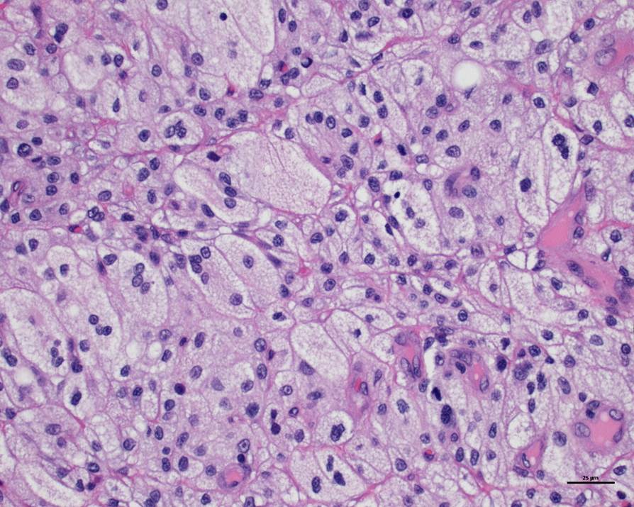
Photomicrograph of a liver biopsy from an acutely photosensitized cow ...
Photomicrograph of Thick Skin Quiz - PurposeGames.com An unregistered player played the game 2 weeks ago About this Quiz This is an online quiz called Photomicrograph of Thick Skin There is a printable worksheet available for download here so you can take the quiz with pen and paper.
Xanthoma: The OTHER Fatty Skin Mass • MSPCA-Angell
09 Histology of skin/How to Draw Thick Skin/Exams Preps ... About Press Copyright Contact us Creators Advertise Developers Terms Privacy Policy & Safety How YouTube works Test new features Press Copyright Contact us Creators ...

Cross Section Thick Skin Tissue Microscopic Stock Photo 203983771 ...
Block1/Fig 10. Dermis of thick skin Fig 10. Dermis of thick skin. This photomicrograph showsthe connective tissue of the skin, referred to as dermis,stained to show the nature and distribution of the elasticfibers (EF), which appear purple. The collagen fibers (CF)have been stained by eosin, and the two fiber types are easilydifferentiated. The elastic fibers of the dermis have a 3Dinterlacing configuration, thus the variety of ...
ANAT2511 Integumentary System - Embryology
photomicrographs of thin skin Flashcards - Quizlet photomicrographics of thick skin. 6 terms. Madison_Tacquard. Figure 6.3 (skin section) 19 terms. Madison_Tacquard. Other sets by this creator. Adult Lab Values. 36 terms. ... Start studying photomicrographs of thin skin. Learn vocabulary, terms, and more with flashcards, games, and other study tools. Home. Subjects. Explanations. Create. Study ...

32 Label The Photomicrograph Of Thick Skin - Labels Database 2020
› pmc › articlesMR imaging of ovarian masses: classification and differential ... Dec 16, 2015 · It is a benign germ cell tumour consisting of at least two of the three embryogenic germ cell layers, and usually contains ectodermal (skin, brain), mesodermal (fat, bone) and/or endodermal (thyroid tissue, gastrointestinal and bronchial epithelium) mature tissue . Simultaneous presence of these components leads to a complex and heterogeneous ...
Stratum lucidum – Definition, Location, Functions and Pictures - Body Terms
› 44502808 › The_Developing_Human(PDF) The Developing Human-Clinically Oriented Embryology by ... Keith L. Moore

Skin care -Skin histology | the dynamic natural skin care
Block1/Fig 11. Hypodermis of the thick skin. Fig 11. Hypodermis of the thick skin. The lower magnification photomicrograph shows part of the hypodermisof the thick skin. It contains abundant adipocytes. Theadipocyte (Ad) nucleus is compressed and displaced to oneside of the stored lipid droplets and the cytoplasm includingorganelles is reduced to a small rim (Fig 11c). Fig 11ashows several adipocytes and nerve fiber bundles (NB).Fig 11b ...

35 Label The Photomicrograph Of Thin Skin. - Labels Information List
Solved The photomicrograph of thick skin | Chegg.com The photomicrograph of thick skin ; Question: The photomicrograph of thick skin . This problem has been solved! See the answer See the answer See the answer done loading. Show transcribed image text Expert Answer. Who are the experts? Experts are tested by Chegg as specialists in their subject area. We review their content and use your feedback ...

mammal trachea cells - Google Search | bio | Pinterest
photomicrograph of the epidermal layer in thick skin ... photomicrograph of the epidermal layer in thick skin Diagram | Quizlet photomicrograph of the epidermal layer in thick skin STUDY Learn Write Test PLAY Match + − Created by abba_dabba_17 Terms in this set (6) stratum corneum ... stratum lucidum ... stratum granulosum ... stratum spinosum ... stratum basale ... dermis ... OTHER SETS BY THIS CREATOR
Post a Comment for "42 photomicrograph of thick skin"